31+ Endometrium Histology Pics

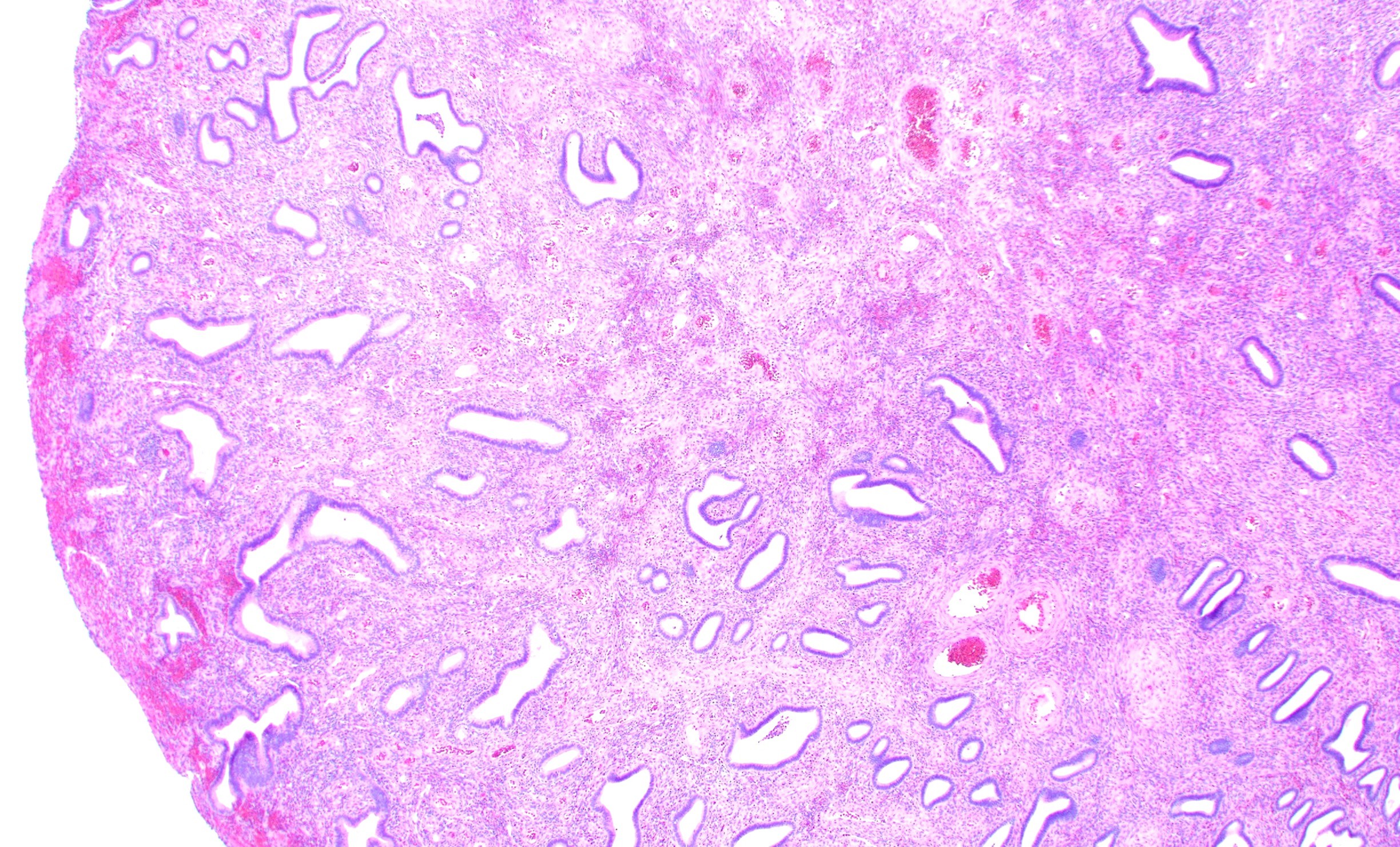
Histology of the uterus · the myometrium (uterine musculature) comprises a complex of three smooth muscle layers which are microscopically difficult to separate . The early secretory endometrium is a stage of the menstrual cycle in which a nearly mature endometrium has a layer of grandular epithelium with round nuclei, thickened endometrium and curled uterine glands with collections of glycogen withi. The uterus is made up of an external layer of smooth muscle called the myometrium, and an internal layer called the endometrium. The top two panels represent examples of benign proliferative a and secretory b human endometrium. Mucosal layer lining the uterine cavity composed of endometrial glands and specialized stroma;

The uterus is made up of an external layer of smooth muscle called the myometrium, and an internal layer called the endometrium. Histology of the uterus · the myometrium (uterine musculature) comprises a complex of three smooth muscle layers which are microscopically difficult to separate . Nicole galan, rn, is a registered nurse and the author of the everything fertility book. jessica shepherd, md, very. Before pathologists can examine tissue and blood samples to uncover abnormalities and diagnose disease. Most specimens are taken because of abnormal uterine . Mucosal layer lining the uterine cavity composed of endometrial glands and specialized stroma; It has a basal layer and a functional layer: The top two panels represent examples of benign proliferative a and secretory b human endometrium.
Before pathologists can examine tissue and blood samples to uncover abnormalities and diagnose disease.
Functional layer of endometrium glands and stroma. Anatomy & histology · endometrium: Nicole galan, rn, is a registered nurse and the author of the everything fertility book. jessica shepherd, md, very. Mucosal layer lining the uterine cavity composed of endometrial glands and specialized stroma; Histology of the uterus · the myometrium (uterine musculature) comprises a complex of three smooth muscle layers which are microscopically difficult to separate . Learn how a secretory endometrial biopsy is performed before you have tissue samples taken from the lining of your uterus. It has a basal layer and a functional layer: The uterus is made up of an external layer of smooth muscle called the myometrium, and an internal layer called the endometrium. Pathology laboratories are central to a hospital or clinic's ability to diagnose illnesses. The early secretory endometrium is a stage of the menstrual cycle in which a nearly mature endometrium has a layer of grandular epithelium with round nuclei, thickened endometrium and curled uterine glands with collections of glycogen withi. Arcuate, radial, straight, and coiled arteries. The simplest classification of these layers is their division into a mucosal layer, or endometrium, a muscularis layer, or myometrium, and a serosal layer, or . The endometrium consists of a simple columnar epithelium, forming numerous tubular glands, supported by a thick vascular stroma.
Functional layer of endometrium glands and stroma. Nicole galan, rn, is a registered nurse and the author of the everything fertility book. jessica shepherd, md, very. Arcuate, radial, straight, and coiled arteries. Anatomy & histology · endometrium: The endometrium is the inner epithelial layer, along with its mucous membrane, of the mammalian uterus.

The endometrium consists of a simple columnar epithelium, forming numerous tubular glands, supported by a thick vascular stroma. Pathology laboratories are central to a hospital or clinic's ability to diagnose illnesses. Arcuate, radial, straight, and coiled arteries. The top two panels represent examples of benign proliferative a and secretory b human endometrium. Functional layer of endometrium glands and stroma. Learn how a secretory endometrial biopsy is performed before you have tissue samples taken from the lining of your uterus. Nicole galan, rn, is a registered nurse and the author of the everything fertility book. jessica shepherd, md, very. The uterus is made up of an external layer of smooth muscle called the myometrium, and an internal layer called the endometrium.
Before pathologists can examine tissue and blood samples to uncover abnormalities and diagnose disease.
The early secretory endometrium is a stage of the menstrual cycle in which a nearly mature endometrium has a layer of grandular epithelium with round nuclei, thickened endometrium and curled uterine glands with collections of glycogen withi. In many histopathology laboratories, endometrial specimens account for a major proportion of the workload. The simplest classification of these layers is their division into a mucosal layer, or endometrium, a muscularis layer, or myometrium, and a serosal layer, or . Mucosal layer lining the uterine cavity composed of endometrial glands and specialized stroma; The uterus is made up of an external layer of smooth muscle called the myometrium, and an internal layer called the endometrium. Anatomy & histology · endometrium: Functional layer of endometrium glands and stroma. Pathology laboratories are central to a hospital or clinic's ability to diagnose illnesses. Arcuate, radial, straight, and coiled arteries. The endometrium is the inner epithelial layer, along with its mucous membrane, of the mammalian uterus. Most specimens are taken because of abnormal uterine . Learn how a secretory endometrial biopsy is performed before you have tissue samples taken from the lining of your uterus. It has a basal layer and a functional layer:
Before pathologists can examine tissue and blood samples to uncover abnormalities and diagnose disease. The simplest classification of these layers is their division into a mucosal layer, or endometrium, a muscularis layer, or myometrium, and a serosal layer, or . Nicole galan, rn, is a registered nurse and the author of the everything fertility book. jessica shepherd, md, very. In many histopathology laboratories, endometrial specimens account for a major proportion of the workload. The endometrium consists of a simple columnar epithelium, forming numerous tubular glands, supported by a thick vascular stroma.

Nicole galan, rn, is a registered nurse and the author of the everything fertility book. jessica shepherd, md, very. The simplest classification of these layers is their division into a mucosal layer, or endometrium, a muscularis layer, or myometrium, and a serosal layer, or . Functional layer of endometrium glands and stroma. Histology of the uterus · the myometrium (uterine musculature) comprises a complex of three smooth muscle layers which are microscopically difficult to separate . Arcuate, radial, straight, and coiled arteries. Learn how a secretory endometrial biopsy is performed before you have tissue samples taken from the lining of your uterus. Anatomy & histology · endometrium: The top two panels represent examples of benign proliferative a and secretory b human endometrium.
Pathology laboratories are central to a hospital or clinic's ability to diagnose illnesses.
The uterus is made up of an external layer of smooth muscle called the myometrium, and an internal layer called the endometrium. Histology of the uterus · the myometrium (uterine musculature) comprises a complex of three smooth muscle layers which are microscopically difficult to separate . Nicole galan, rn, is a registered nurse and the author of the everything fertility book. jessica shepherd, md, very. Before pathologists can examine tissue and blood samples to uncover abnormalities and diagnose disease. Mucosal layer lining the uterine cavity composed of endometrial glands and specialized stroma; Arcuate, radial, straight, and coiled arteries. In many histopathology laboratories, endometrial specimens account for a major proportion of the workload. The endometrium is the inner epithelial layer, along with its mucous membrane, of the mammalian uterus. It has a basal layer and a functional layer: Anatomy & histology · endometrium: Most specimens are taken because of abnormal uterine . The simplest classification of these layers is their division into a mucosal layer, or endometrium, a muscularis layer, or myometrium, and a serosal layer, or . The top two panels represent examples of benign proliferative a and secretory b human endometrium.
31+ Endometrium Histology Pics. Mucosal layer lining the uterine cavity composed of endometrial glands and specialized stroma; The top two panels represent examples of benign proliferative a and secretory b human endometrium. Functional layer of endometrium glands and stroma. The uterus is made up of an external layer of smooth muscle called the myometrium, and an internal layer called the endometrium. The endometrium consists of a simple columnar epithelium, forming numerous tubular glands, supported by a thick vascular stroma.
Arcuate, radial, straight, and coiled arteries endometrium. The top two panels represent examples of benign proliferative a and secretory b human endometrium.


