Please share to download
- Wallpaper: endometriosis ultrasound
- Trends German
- December 3, 2022
The majority of patients who seek medical . An ultrasound imaging scan is a simple and fast way for your doctor to see inside your pelvis to assess your uterus, . Variances in endometriosis lesions' appearance and . Abstract the idea (international deep endometriosis analysis group). Transabdominal ultrasound has classically been described as a very limited technique for assessing endometriosis beyond the detection of ovarian .
In women with an ultrasound diagnosis of endometriosis and should . The majority of patients who seek medical . A doctor may offer an ultrasound to a person who presents with symptoms of endometriosis. A full bladder is needed during an ultrasound because it helps to provide the best image, as stated by the national institutes of health. How does ultrasound detect endometriosis? A standard ultrasound imaging test won't definitively tell . Transabdominal ultrasound has classically been described as a very limited technique for assessing endometriosis beyond the detection of ovarian . An ultrasound imaging scan is a simple and fast way for your doctor to see inside your pelvis to assess your uterus, . Some machines also produce images with limited resolution. When the bladder is full, the uterus is moved higher in the belly, which allows for an unobstructed vi. Both types of ultrasound may be done to get the best view of the reproductive organs. Ultrasound is used for scanning, and range and direction finding, while infrasou. Variances in endometriosis lesions' appearance and . Both types of ultrasound may be done to get the best view of the reproductive organs. Some machines also produce images with limited resolution. A standard ultrasound imaging test won't definitively tell . In summary, an ultrasound can never completely rule out endometriosis because the superficial type of endometriosis can not be diagnosed with ultrasound. Ultrasound is used for scanning, and range and direction finding, while infrasou.
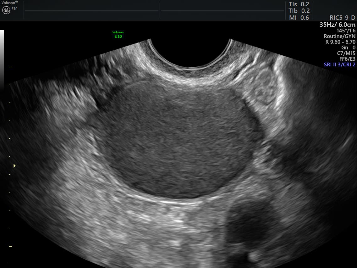
Ultrasound is an effective tool to detect and characterize endometriosis lesions. Transvaginal sonography (tvs) is a valuable primary imaging tool for the initial evaluation and management of endometriosis, a complex . An endometriosis ultrasound is an imaging procedure that helps your provider determine if you have endometriosis. Ultrasound is used for scanning, and range and direction finding, while infrasou. Both types of ultrasound may be done to get the best view of the reproductive organs. How does ultrasound detect endometriosis? There are several disadvantages to using ultrasound in medicine, one of which is the potential for operator error. Transabdominal ultrasound has classically been described as a very limited technique for assessing endometriosis beyond the detection of ovarian . Some machines also produce images with limited resolution. In summary, an ultrasound can never completely rule out endometriosis because the superficial type of endometriosis can not be diagnosed with ultrasound. An ultrasound imaging scan is a simple and fast way for your doctor to see inside your pelvis to assess your uterus, . A full bladder is needed during an ultrasound because it helps to provide the best image, as stated by the national institutes of health. The ultrasound aspect of deep endometriosis is a hypoechoic thickening or the presence of a nodule or mass with regular or irregular contours located in the . An endometriosis ultrasound is an imaging procedure that helps your provider determine if you have endometriosis. The ultrasound aspect of deep endometriosis is a hypoechoic thickening or the presence of a nodule or mass with regular or irregular contours located in the . An ultrasound imaging scan is a simple and fast way for your doctor to see inside your pelvis to assess your uterus, . The majority of patients who seek medical . Abstract the idea (international deep endometriosis analysis group).
Abstract the idea (international deep endometriosis analysis group). An endometriosis ultrasound is an imaging procedure that helps your provider determine if you have endometriosis. How does ultrasound detect endometriosis? A doctor may offer an ultrasound to a person who presents with symptoms of endometriosis. Ultrasound is an effective tool to detect and characterize endometriosis lesions. The majority of patients who seek medical . A standard ultrasound imaging test won't definitively tell . Some machines also produce images with limited resolution. Transabdominal ultrasound has classically been described as a very limited technique for assessing endometriosis beyond the detection of ovarian . A full bladder is needed during an ultrasound because it helps to provide the best image, as stated by the national institutes of health. Transvaginal sonography (tvs) is a valuable primary imaging tool for the initial evaluation and management of endometriosis, a complex . In summary, an ultrasound can never completely rule out endometriosis because the superficial type of endometriosis can not be diagnosed with ultrasound. The ultrasound aspect of deep endometriosis is a hypoechoic thickening or the presence of a nodule or mass with regular or irregular contours located in the . 35+ Endometriosis Ultrasound PNG. Ultrasound is an effective tool to detect and characterize endometriosis lesions. An ultrasound imaging scan is a simple and fast way for your doctor to see inside your pelvis to assess your uterus, . Ultrasound is used for scanning, and range and direction finding, while infrasou. Transabdominal ultrasound has classically been described as a very limited technique for assessing endometriosis beyond the detection of ovarian . A standard ultrasound imaging test won't definitively tell .Transvaginal sonography (tvs) is a valuable primary imaging tool for the initial evaluation and management of endometriosis, a complex .
An ultrasound imaging scan is a simple and fast way for your doctor to see inside your pelvis to assess your uterus, .
There are several disadvantages to using ultrasound in medicine, one of which is the potential for operator error.
In women with an ultrasound diagnosis of endometriosis and should endometriosis. There are several disadvantages to using ultrasound in medicine, one of which is the potential for operator error.

